Close
0%
0%

DIY Space Grade DNA Analyzer

Detect DNA non optically by measuring the electrical properties of analytes as they pass by a detector

Similar projects worth following
In 1948 Arne Tiselius was awarded the Nobel Prize in chemistry for his work in electrophoresis. The genetic code is now an open book that would otherwise still be closed. This project makes use of the LTCC devices covered in the ongoing "DIY Space Grade PCBs" for detecting DNA. A microfluidic signal processor is constructed using the LTCC as a substrate that support the electrodes used in the sensor. Specifically a set of two silver/silver chloride electrodes are energized via an H-switch and two additional electrodes are used as detectors across the fluid channel to measure changes in impedance.

Those aspects of the detector and it's fabrication that relate directly as part of the LTCC technology will be covered in the PCB project. Those that relate to the use of PDMS for example will be covered within this project.

Let's get started!

In the processing of DNA, RNA, proteins and all the other parts of a living organism, for which we as scientists concern ourselves there is frequently a need to move or process the materials under study. Lets suppose one wishes to digest some DNA using a restriction enzyme. Typically, some DNA would be pipetted into some appropriately sized container, and another bit of enzyme pipetted from another container and the two mixed, etc. Then after awhile, the results of the reaction are pipetted into a well of a gel made up of agarose or an acrylic "PAGE", and the gel is "run" using a high voltage. 400 Volts is certainly typical.

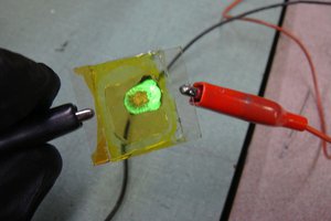
While many might view electrophoresis as a mechanism for sorting out DNA it is more fundamentally a "prime mover". It's as if a river is flowing an you can float your boat and let the current take you down stream. If you can steer your inner tube, canoe, row boat, or yacht, then can take it to the ocean if that's your objective.

This project is about making a rudder that allows one direct electrophoretic flow in a controlled direction. The first step is to reliably detect what's in the water. The problem is of course that's there a lot more in the river than what's on the surface. There can be no satisfaction in only being able to detect just canoes. If this part of the river can't sort out the fish from the algae, there is another section down stream, two locks left, and one tributary right expressly designed for that function.

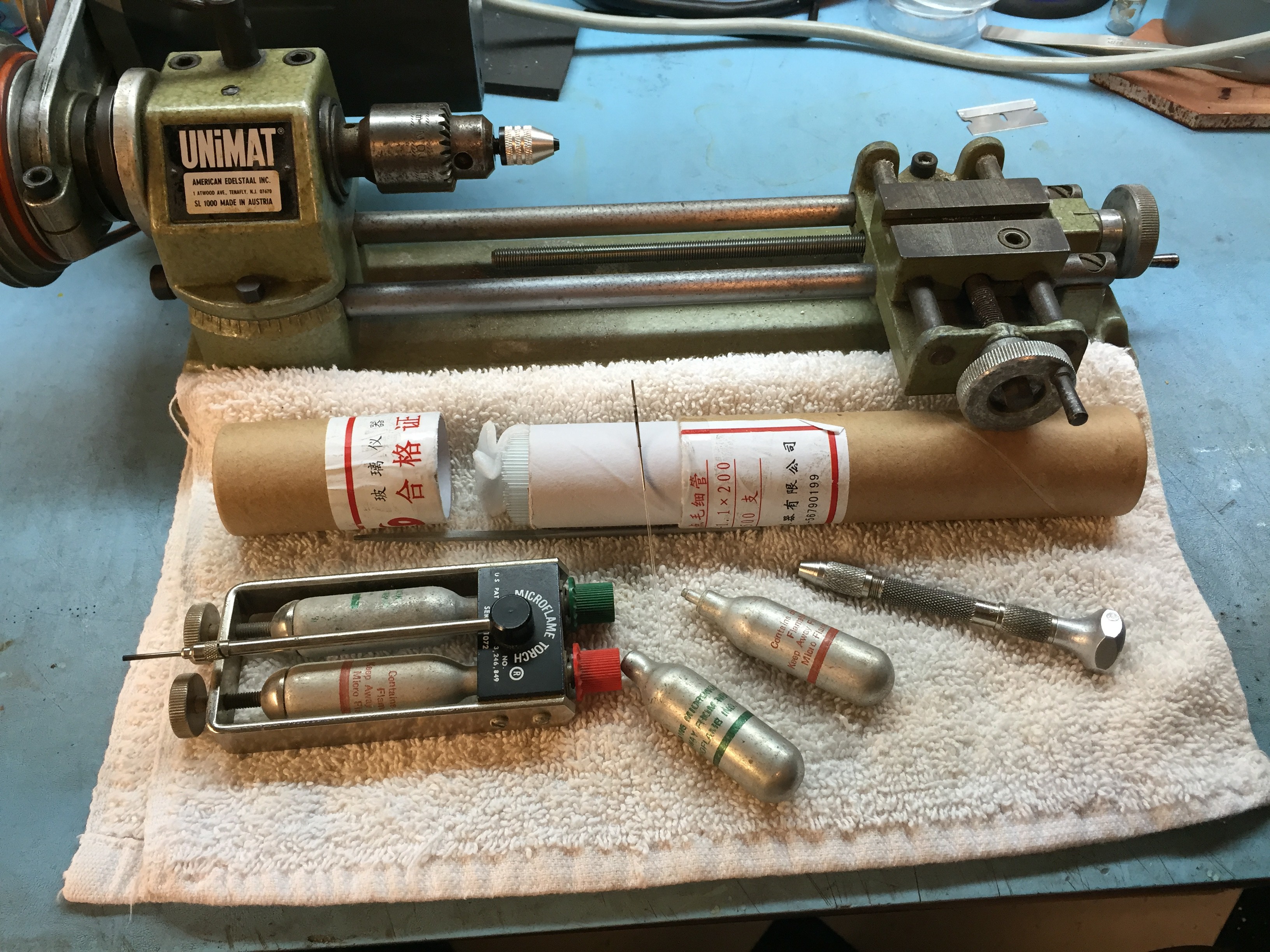
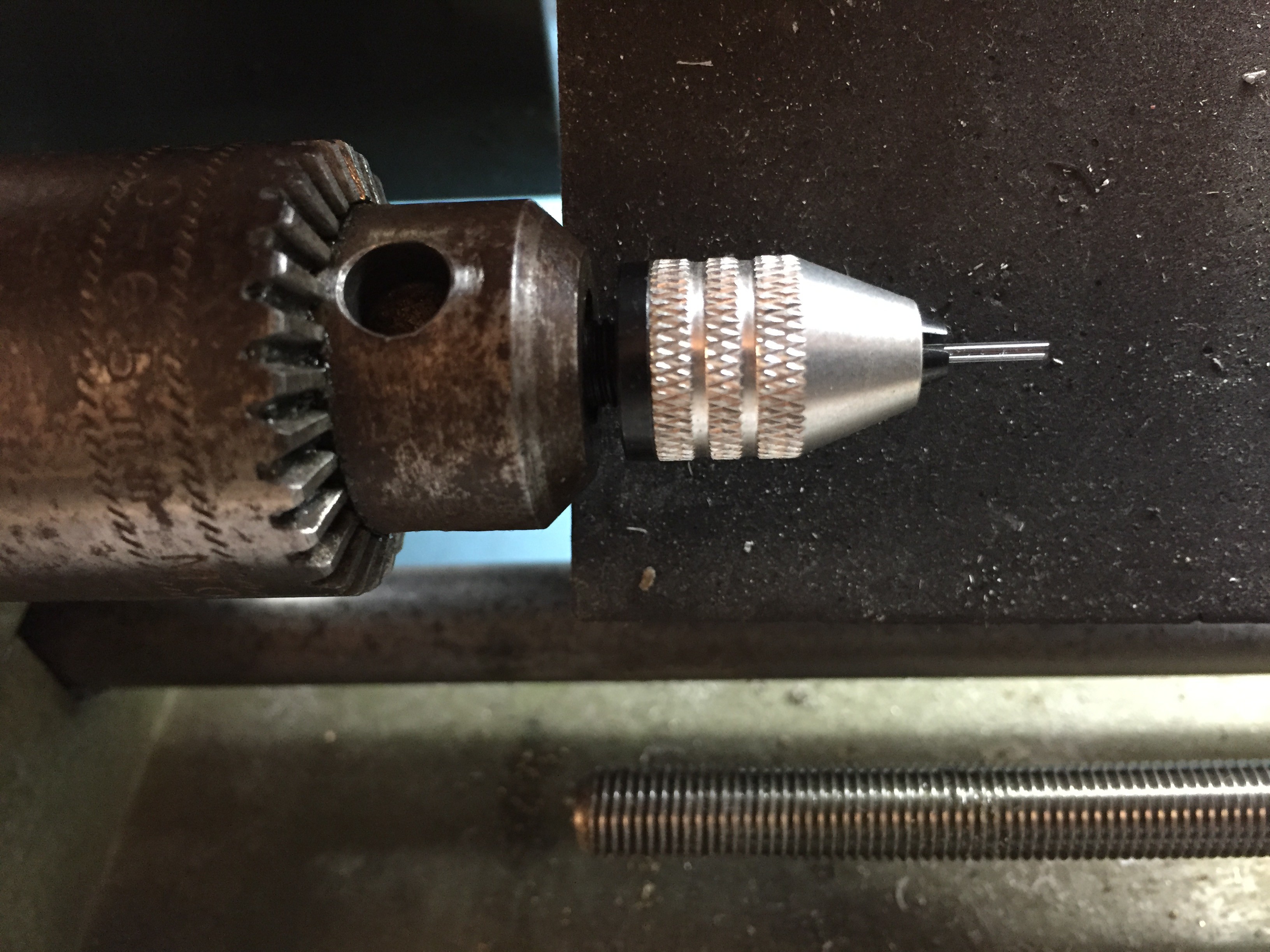
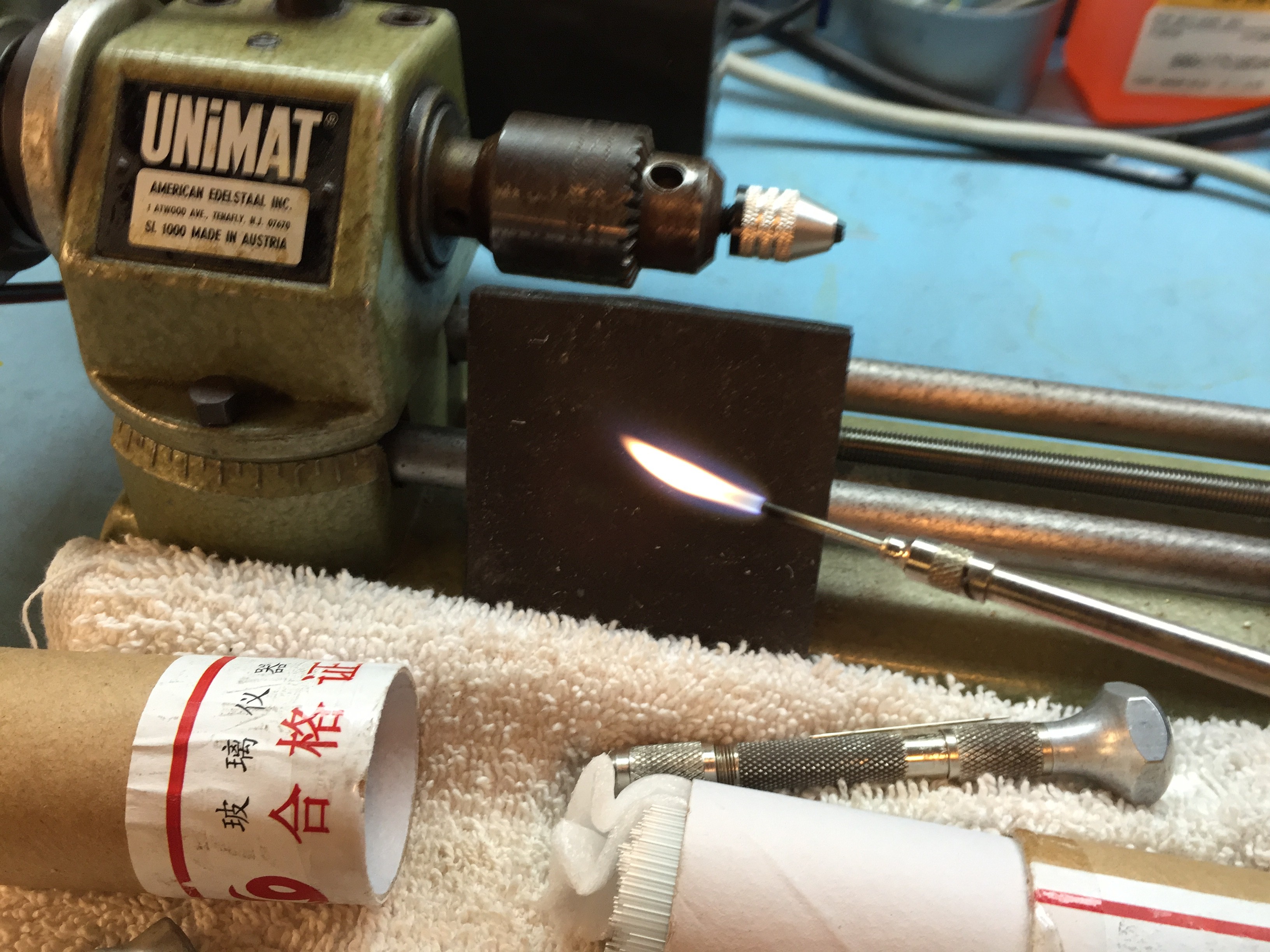
From an engineering perspective the view is a bit different. Normally, electronics and water don't mix. Even worse, biological fluids and those fluids used to process biologicals are loaded with salts. Further, the electronics and fluids are by necessity intimate.

There are a vast collection of physical effects that may be exploited for the detection of substances. So many that we may become overwhelmed by our options. Our design options are greatly expanded however if we utilize methods that by default are immune to the effects of water, salts, and basic incompatibilities inherent in conventional technologies. There is of course on technology that offers a solution. It is called LTCC.

  • 1 × PDMS Poly dimethylsiloxane
  • 1 × DNA Ladder A collection of DNA fragments of different lengths
  • 1 × Electrophoresis buffer Water with a collection of various compounds designed to aid in the flow of materials
  • 1 × Capillary tubing Hollow glass tubing, in this case the inner bore is 1mm
  • 1 × H-Switch A simple electrical switch that either sinks or sources a flow of current

View all 8 components

  • Follow up on yesterdays Agarose/PDMS experiment

    Chuck Glasser07/11/2016 at 23:22 0 comments

    I know for some it might be a bit underwhelming to see a few picture taken of a piece of silicon rubber with what appears to be an image of a few lines in relief. However for others it may well be the freakin holy grail! So show a little respect:)

    Amazing how little bits of dust get picked up, seemingly out of nowhere. You'll note that from the previous log the parts of the line that were the darkest blue, are now the darkest black which suggest those sections are the thickest. I'll have to add a profilometer to the list of instruments that need to be built. I would think, that the depth of the relief is no more than a few microns.

    Incidently, the PDMS pealed off the alumina substrate easily. Perhaps it wasn't clean enough. Like most things made with silicone compounds, not much, including itself sticks to it. If it isn't in the first pour, and you don't have a plasma ozone etcher, then "Forget about it".

  • Off to a good start with a simple, spectacularly successful experiment

    Chuck Glasser07/10/2016 at 21:36 0 comments

    If you are interested in Microfluidics the Princeton Microfluidics Laboratory is a great place to start. No matter where your reading may take you you will find numerous reference to the use of SU-8 as a way of making master molds for the casting of PDMS, the preferred material for making microfluidic devices. While, I'm sure the use of SU-8 is a fantastic method, it's to my way of thinking, too much technology for a simple problem, especially in consideration of the resources required to implement the technique.

    Briefly, microchannel paths are drawn using a calligraphy pen using molten agarose as the ink and PDMS is cast and cured directly over the drawing producing a microfluidic channel.

    The purpose of the Space Grade experiments and machine development in support of the effort are to explore what is possible using a ceramic as a substrate for projects in Molecular Biology and Neuro Interposers and Neuro Cybernetic devices. While Molecular Biology as a term needs no explanation the term Neural Interposer does. An interposer is a electrical device for routing one connection to another. For a prosthetic device a neural interposer forms a bridge between what may be measured electrically in terms of muscle and nerve activity at the stump to the prosthetic device designed replace some or all of the missing function. Although based upon entirely different principles than are found in a prosthetic limb, a "Brain Computer Interface" is also a neural interposer.

    The devices all slightly larger than a poker chip have a hexagonal shape and are called tokens. On one side of the the token may be found electrodes that are designed to interface to fluids. On the other side the electronics that generate or process the signals at the electrodes.

    For tokens to be successful they must respond to the problems of interfacing with fluids as the duck takes to water. Similarly, the electronics located within the very same device must be immune to both water and the corrosive salts that will always be present. Interesting problem!

    This test concerned itself with the immiscibility of agarose and PDMS.

    50 uL of Cyan inkjet ink was mixed with 50 uL of 0.8% agarose and using a calligraphy nib drawn onto an alumina substrate. The fluid was kept warm in a water bath to keep the agarose from gelling on the pin. There is no significance to the choice of symbols.

    5.5 mL of Sylgard 184 PDMS was then mixed and poured directly on top of the agarose ink and cured in a lab oven at 80 degrees C for one hour.

    under the heat of the curing process the PDMS continued to spread across the surface of the substrate. Note that there is absolutely no evidence of smearing or diffusion of the ink into the PDMS 50 mil thick. This observation strongly suggest that the two reagents are indeed imiscible.

    What this means is that, in this case, microcapillaries may be drawn directly on a ceramic substrate and that the PDMS may be cast directly over the agarose channels, without effect.

    This alone however will not be sufficient. DNA is known to have a extremely high affinity for glass. The same should be expected for alumina and composite ceramics such as cofired ceramics that are made up of almost equal amounts of alumina and glass. It will be necessary to passivate the substrate surface using a surfactant such as bovine serum albumin or polyethylene glycol. Ideally, it may be effective to draw the albumin pattern on a layer of PDMS that has undergone activation via ozone. By this mechanism one could avoid passivation.

    Each of these avenues needs to be explored. Ultimately, after all the experiments are performed it may happen that SU-8 is still the champion. Only time, experiments, and a working LTCC process will tell the tale.

  • Adding a flange to a capillary tube

    Chuck Glasser07/10/2016 at 17:02 0 comments

    Reality is a cruel taskmaster. Making a transition from tiny tubing to a planar structure like a Lab on a Chip is a perfect example.

    There is a very old saying in electronics, especially where it concerns debugging something that was once working but now mysteriously is not functioning, "99% of all problems are at the interface". Nothing could more true in regards to microfluidics and Lab on a Chip. After having gone to enormous trouble preparing a substrate, laying down gold, silver and other precious metals, spending all sorts of time on artwork, generating cut files and G-Code, miking up countless batches of PDMS, preparing reagents, ordering materials, and much, much more. We finally come to the central problem.

    How exactly am I to get 10 uL of this DNA into that tiny, little, thin sliver of a machine? Along with the running buffers and all the other fluids involved, there might be lots of fluid connections. Fluids here includes any gases like air needed for pneumatic valves. One common way would be to mount all the tubing interfaces in a manifold.

    One simple and effective way join a tube made of vinyl, silicone, or polyamide is to slip it over the end of a glass capillary tube that is flanged. Then, one simply seals the connection with an adhesive or clamp to the substrate and the task is done.

    This is the equipment I used about 40 years ago when I developed the technique. I was in for a few surprises.

    The basic tools are a glass lathe, capillary tubing, Oxy/Butane torch, and pin vice holding a needle.

    So spin up the capillary in the lathe.

    Heat the end of the capillary with a tiny hot flame while using the pin vise to keep the opening from closing, If you get the timing just right , you get a flange. You'll have to trust me on this one. It works.

    As I mentioned, this glass working discovery was made quite some time ago. Between the then and now, Microflame has gone out of business. All of my spare oxygen cartridges have gone empty, and the walls of the capillary tubing that I recently purchased are too thin. Oh crap!

    I promise to update this log with present day tools and materials that work! But it's going to take a few weeks while new material arrives from the other side of the world.

    I even promise a nice video illustrating the technique.

  • Use a PCB editor to define fluid paths

    Chuck Glasser07/09/2016 at 16:03 0 comments

    Molecular Biology is certainly different than a discipline like electrical engineering. One of the more obvious examples are the consumables. In general, things are always being added to little containers, like say a restriction enzyme. After the reaction, the restriction enzyme doesn't go back up on the self, it's consumed. Or let's say, the experiment is to identify a protein using two dimensional electrophoresis. The protein of interest is identified spatially, cut out of the gel, dropped into a tiny vile, and the rest of the gel dumped in the trash.

    Suppose instead that the goal is to make a large microfluidic signal processor in which all the operations of mixing fluids, adding various proteins, and performing separation process occur within a machine.

    If one wishes to go about building a device in which the build of the operations occur in a very small space, "Lab on a Chip", then it will be necessary to generate artworks that define the physical pathways, electrodes, reservoirs, fluid connections, and components, that make up that device.

    As mentioned earlier the substrate for the devices will be constructed on a ceramic. The electrodes will be made of silver. For the fluid pathways, there is really only one choice, polydimethylsiloxane (PDMS). PDMS has a very low surface energy. What that means is that nothing sticks to it. It sticks to itself when it is cast from a liquid, but once it cures it pretty much won't stick to anything, including itself. So if a shape is cast, cured, and another layer cast on top of the first layer, don't expect the two to be one integral body. If it is necessary to join two layers, it is necessary to etch the first with ozone to prepare the surface. Once prepared, the is only a very sort period of time, according to the literature, no more than 20 minutes, before the second layer must be poured.

    Because PDMS is an elastomer it does make a great mechanical seal.

    So, where exactly does the pattern of fluid pathways come from? Is it a positive photo mask system or a negative one? Are the pathways made by first patterning the desired shape in another material like SU8 or are they made by xurography, Latin for a cutting plotter?

    No matter the polarity, may I recommend the open source triumph, Kicad. As one of Kicads attributes is the ability to export SVG files. From there, one may import a SVG graphic into another open source wonder, Inkscape. From Inkscape it is an easy path to generate G-code for a CNC machine, or HPGL for a graphic plotter, And there you have it. Tremendous power at your fingertips.

    Be prepared for some serious study, Kicad and Inkscape are seriously proficient programs that require practice.

  • Murphy joined me for lunch today

    Chuck Glasser07/04/2016 at 04:34 0 comments

    I was the main course. I wish that guy over at the LTCC PCB shop would fix his press!

    According to the latest search, circa July 3, 2016, it cost around $1000 to process the human genome with 30X coverage, meaning that there is an error around .1%. So, every 1 out of 1000 base pairs there will be an error. A C should have been a T, a A a G, etc.. Absolutely, of no relevance. Science has only one direction, forward.

    So, what exactly does Space Grade mean? When, in a few years, SpaceX lands a crew on Mars, they will leave behind at least one instrument for molecular biology. Thousands of years later, archaeologist rediscover the landing site and find in the sand, half buried, lies a DNA analyser, still functional, that stands as testament to the best engineering principles.

    If we are to build instruments that will endure, then we must build them from ceramics as that is the only material that is evidenced to survive the passage of time.

    Machines that last. Based upon first principals that endure the passage of time. How does one go about building such machines?

    Had this been a journal paper the title would be, "DNA Analysis by Restriction Enzyme Mapping". If you take a DNA molecule and digest it through the application of restriction enzymes, then it will be cut into a collection of smaller fragments. The fragments will be defined by the distance between digestion sites. If the fragments are then electrophoresed, they will be sorted by their molecular size. So, as each fragment passes by a detector, they may be detected, and in the words of Feyman to paraphrase, since I haven't identified the exact quote, "them their electrons that go to the left and those that go to the right". If you can detect it, then you can switch it. And repeat the process. As each chain of nucleotides is digested, producing smaller and smaller chains, the ends of the molecule are defined, until the molecules is of sufficient size to be economically digested via a Sanger process or something like it. The end result is of course a completely sequenced molecule. Run it through again with a different order of restriction to insure correct coverage.

    The purpose of this study is to demonstrate that it is possible to detect DNA via relatively simple instrumentation processes. Thus, having demonstrated that detection is possible, the second problem is to do it on an industrial scale. In a simple proof of concept demonstration one might expect to dissipate a few milliwatts of power in moving DNA from point A to B along with detection. This typically would involve electrophoresis voltages of no more than 20 Volts. In an industrial application one should expect to use upwards of 10 KW, with drive voltages of around 10 KV. Huge difference. At an industrial scale the DNA analysis system is now spread out over a large surface, let's say an ice rink. Thousands of slicers and dicers, digesting, sorting, switching DNA in a constant flow with a new genome entering the system every 30 seconds. For an ice rink area the power dissipation could easily be 1 Mega Watt. The use of an ice rink is deliberate. It is necessary, because of Arhenius, to keep chemical processes at a constant temperature, or at least controllable temperature. Running open loop is verboten!

    Keep in mind that because the process is running at an extremely high operating voltage in the presence of corona. The detectors, never the less, must detect signals at a nano volt level. Giving the overall system dynamic range of around +160 to +200 dB. No system, to this authors knowledge with this dynamic range presently exists! Further, all the communications processes that exist behind the detector must also be capable of operating at a very high common mode. An yet all of this is possible and practical, based upon a device no larger than a poker chip.

View all 5 project logs

Enjoy this project?

Share

Discussions

Similar Projects

Does this project spark your interest?

Become a member to follow this project and never miss any updates