 Myrijam
For the development of our prototype we recreated some experiments from the specialist literature in order to test whether our ideas can be realized. This includes e.g. the attempt to distinguish venous from arterial blood by means of a spectral analysis or the illumination of the tissue with different wavelengths and the recording of the light scatter with one of the camera systems.
During the development phase of the device, we were faced with the question whether there is a discernible difference between arterial and venous blood when illuminating with infrared light.
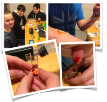
To find this out, we conducted a spectral analysis with diluted blood. We have extracted Venous blood by a venipuncture, but we have not been able to determine arterial blood exactly, but poked a needle into the blood vessels in the finger and thus obtained a mixture of venous and arterial blood.
We found out that there was only a weakly visible difference in the infrared range of the absorption spectra of the blood samples - possibly due to the mixture in the second sample (blood vessels in the finger). Nevertheless, the difference is recognizable and meaningful in the overall representation of the spectrum: The blue absorption curve shows the venous blood, which absorbs the spectral range significantly above approximately 600 nm (orange). This difference, which the spectrometer could only image up to about 900nm, is to be amplified by the image calculation by software. To this end, we have also carried out several tests - both in the visible and in the NIR range.
In this experiment, we examined the sensitivity of the camera for different wavelengths and the reflection of skin and mesh on the inner side of the forearm. For this purpose we have illuminated the skin areas with various colored LEDs - from blue to green, yellow, orange to red - and this "lighting" with the Raspberry Pi camera. We did not use the webcam for this experiment, because it was already equipped with a filter, which only allows infrared light to pass through and almost blocks the visible area. We also wanted to know with which wavelengths in connection with the camera the best results possible can be achieved.
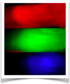
An intensive red illumination is suitable for representing the darker shadows of the veins. However, differences can also be seen in green lighting, which, however, are significantly larger in the case of red LEDs. For the evaluation of the IR illumination, however, a further unknown quantity also plays a role, namely the sensitivity of the camera sensor outside the visible range.
Another method of venous discovery was found in the publication of Asrar / Al-Habaibeh et al. (2016). After we had done first experiments with visible and IR light. This method is used to visualize veins using a thermal imaging camera. The affected site must be cooled before carrying out the procedure. Since the veins transport warm blood into the cooled region, the skin parts, where the large veins are seated, warm up first. This heating process should be able to be imaged by the thermal imaging camera and the veins are so clearly defined by the surrounding structures. We followed the experiment with a thermal imaging camera to check how good the application is with a relatively cheap IR camera.
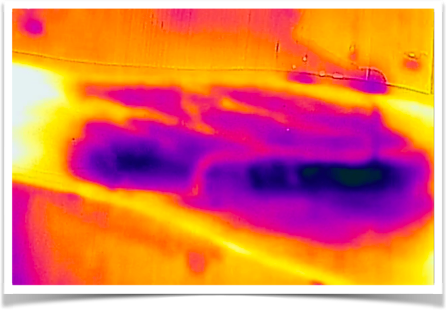
For this, we cooled the arm once with ice cubes and in another run with an icing spray and the warming with the thermal imaging camera recorded as video. In the course of time, the thermal image shows how skin-like areas of the skin heat up - here, veins are recognizable. The surrounding skin areas heat up significantly slower. However, the image and thermal resolution of the camera (FLIRone) used is not particularly high and is 160 × 120 pixels with a thermal sensitivity of 0.1 ° C. This means that no finely resolved structures are recognizable. This could be changed by choosing a model with a larger spatial and thermal resolution. The drawback of this type of method is the enormous cost for thermal imaging cameras with high resolution (several thousand euros); For this reason we do not pursue this procedure any further. In addition, this can be unpleasant with cooling with ice cubes, even with a cold spray, and in the worst case can lead to burns of the skin.
Discussions
Become a Hackaday.io Member
Create an account to leave a comment. Already have an account? Log In.