David H Haffner Sr
-
1 Step Closer To Proving My Design Prototype DAV5 V3 Raman Spectrometer Log#39
12/31/2016 at 16:52 • 0 comments*Addendum to the data below;
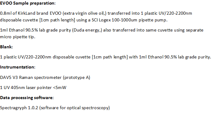
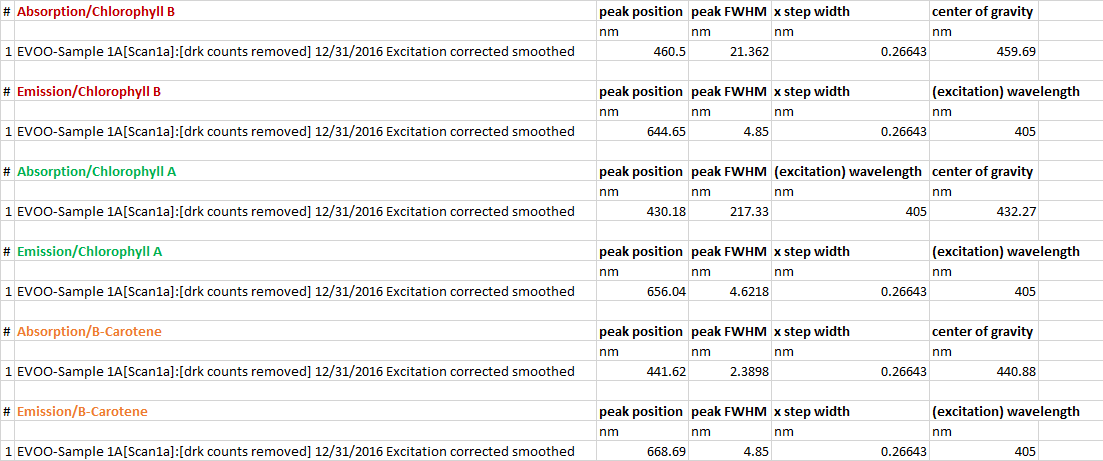
![]() *Abstract, Before continuing any further in phase II testing, I wanted to see just where I am in both optical resolution and spatial resolving power. So I decided to take some spectral data from an EVOO sample I've had for awhile (preserved in a plastic UV cuvette W/an air tight top,) it's about 4 months old and some degradation does show up in the data but it was still an excellent sample to use.
*Abstract, Before continuing any further in phase II testing, I wanted to see just where I am in both optical resolution and spatial resolving power. So I decided to take some spectral data from an EVOO sample I've had for awhile (preserved in a plastic UV cuvette W/an air tight top,) it's about 4 months old and some degradation does show up in the data but it was still an excellent sample to use.The methodology that I am using for this project may seem slow but it's purpose is both methodical and purposeful, in that, every small successful step brings the entire project closer to its main objective, which is, Raman spectroscopy in the 532nm range. Phase III begins when I feel confident enough in the data that, both instrumentally and methodology are sufficient for Raman spectroscopy.
![]()
![]()
![]()
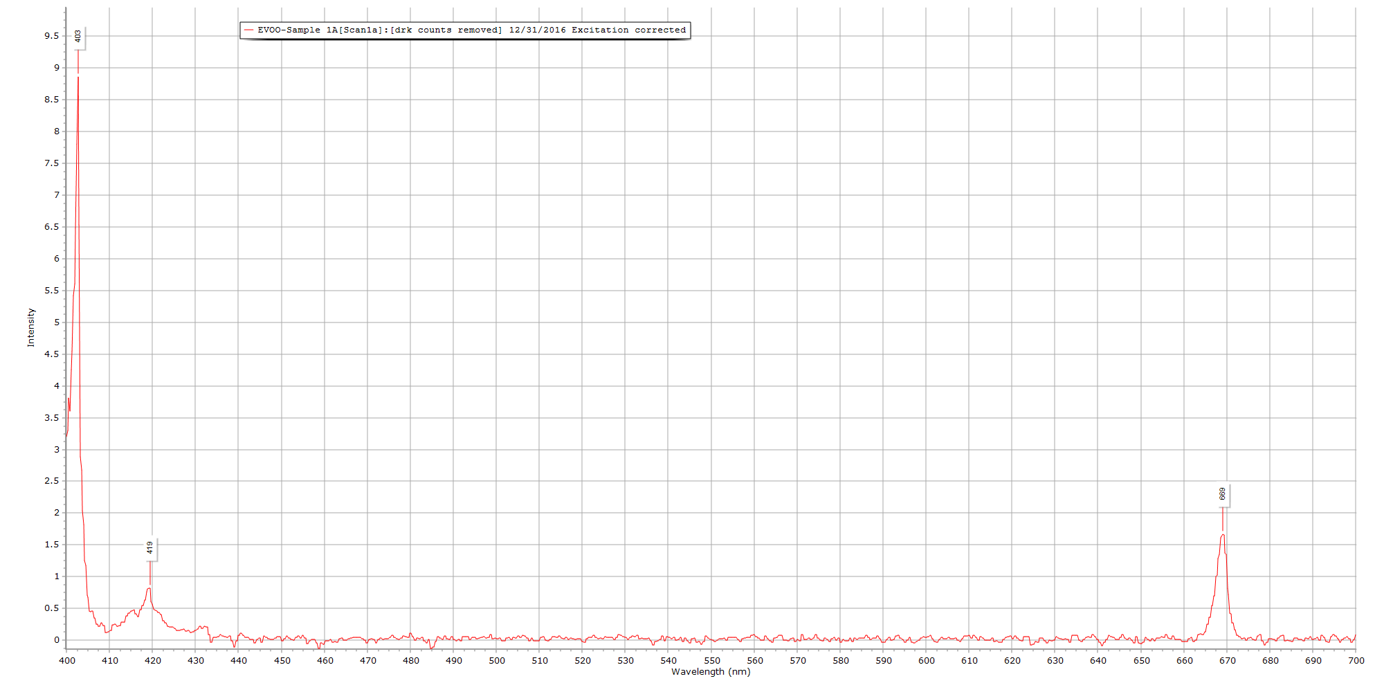
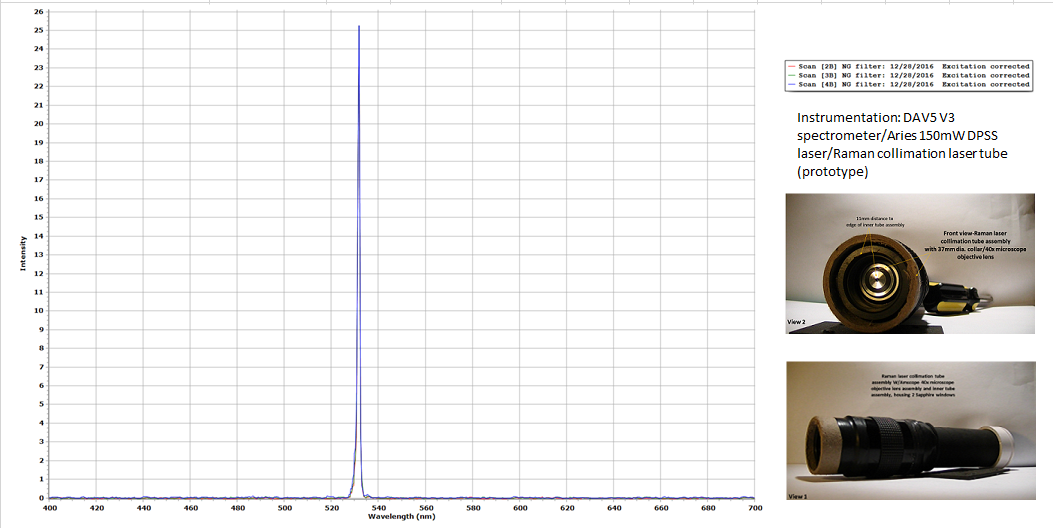
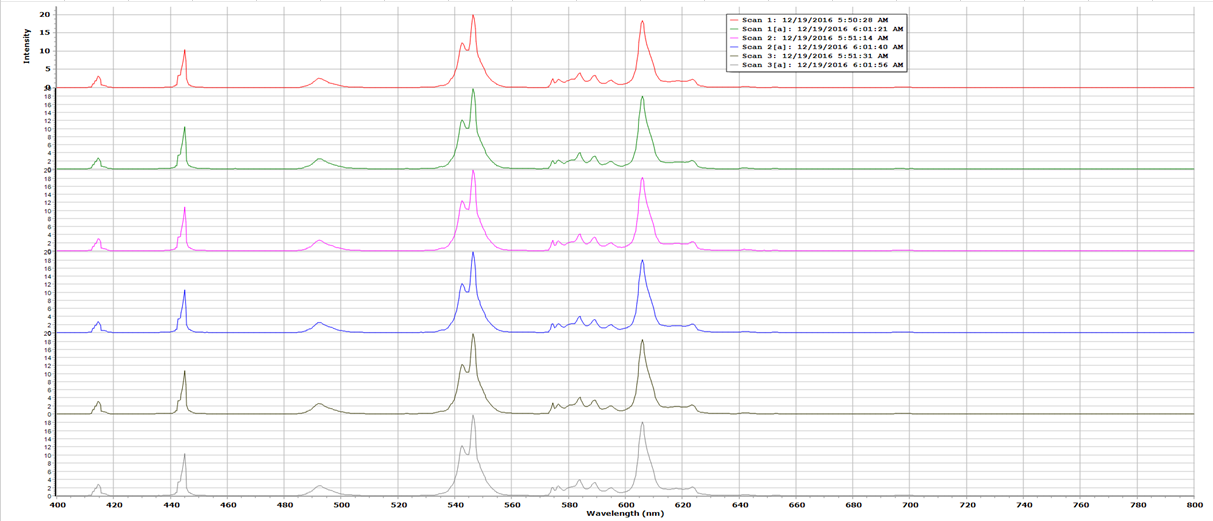
The plot below is the "raw" data:
![]()
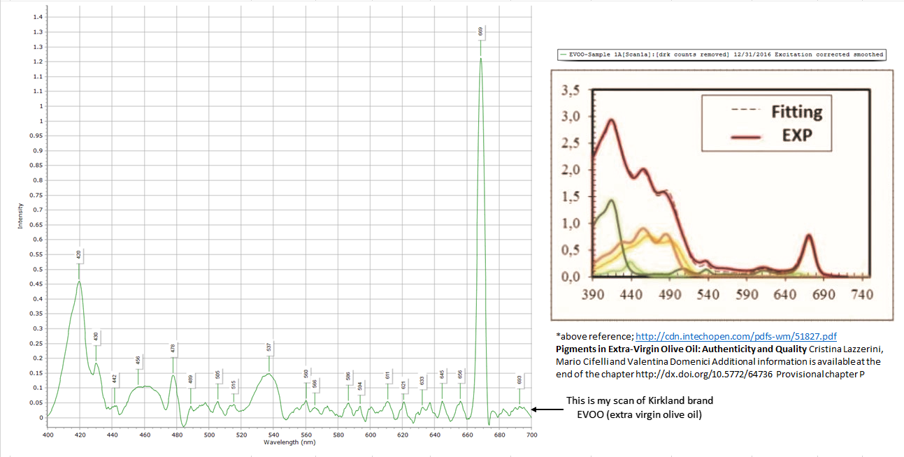
References used:
http://cdn.intechopen.com/pdfs-wm/51827.pdf
Pigments in Extra‐Virgin Olive Oil: Authenticity and Quality Cristina Lazzerini, Mario Cifelli and Valentina Domenici Additional information is available at the end of the chapter
http://dx.doi.org/10.5772/64736 Provisional chapter P
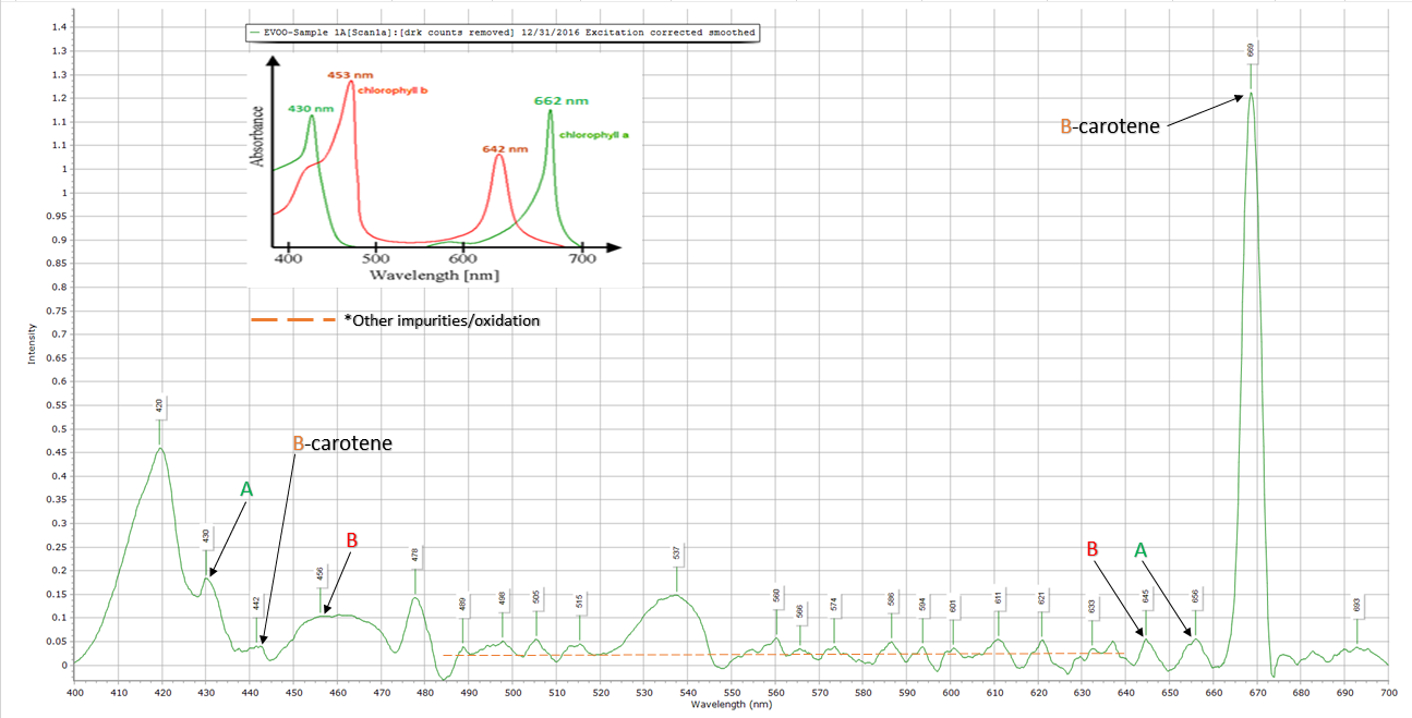
http://www.art-xy.com/2013/03/isolation-of-chlorophyll-and-beta.html - Chlorophyll study reference
-
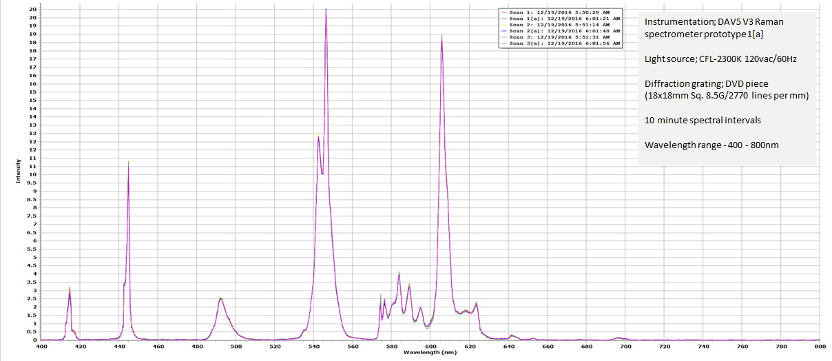
Near Perfect CFL Calibration (2300K/13W/compact fluorescent light)
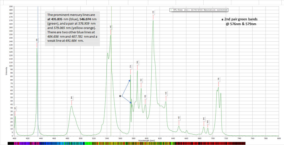
12/31/2016 at 09:13 • 0 commentsProject log#38 [DAV5 V3 Raman spectrometer]; A successful CFL calibration with a high quality "fit" above 95%, my spectral plot data indicates 434nm at the second blue band and 545nm at the first green band (these are the 2 most prominent mercury lines .)
![]()
![]()
This data has an optical resolution of; 0.29nm using the equation:
∆λFWHM =0.84λ/Gwbeam
![]()
-
First Absorption/Emission Testing for The DAV5 V3 Spectrometer Log#37
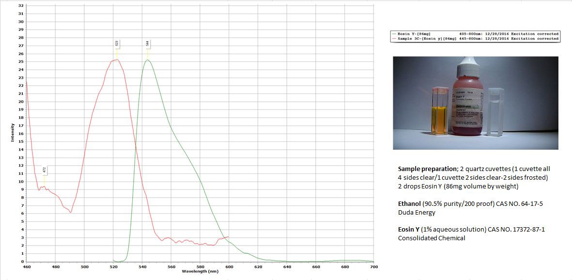
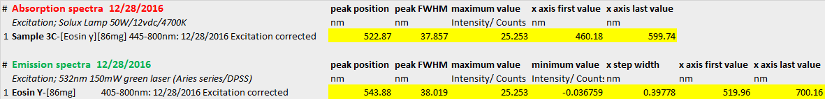
12/29/2016 at 12:11 • 0 commentsProcessing software; Spectragryph 1.02 Instrumentation; DAV5 V3 Raman Spectrometer
![]()
![]()
-
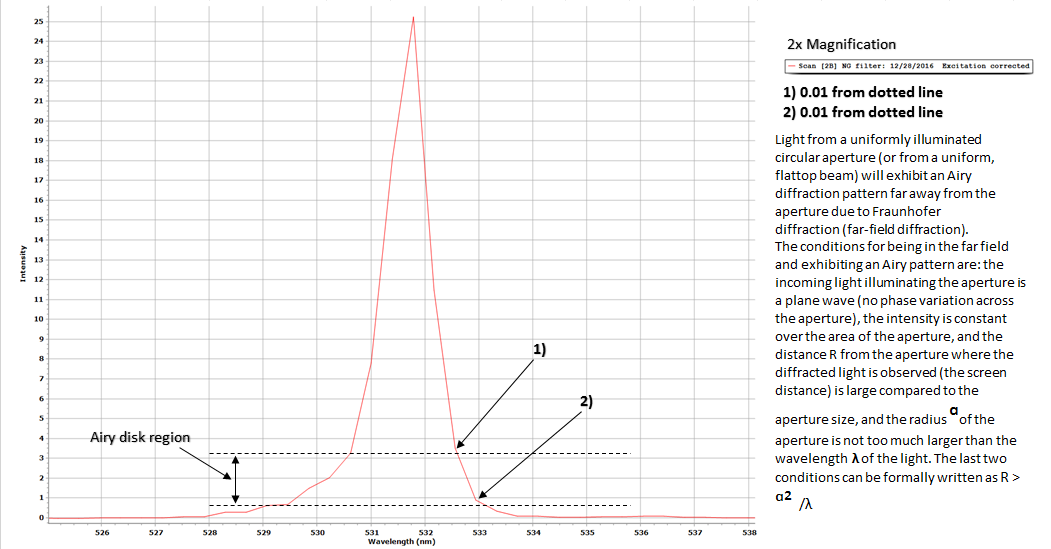
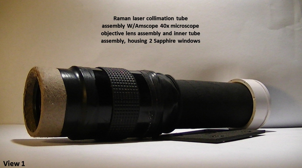
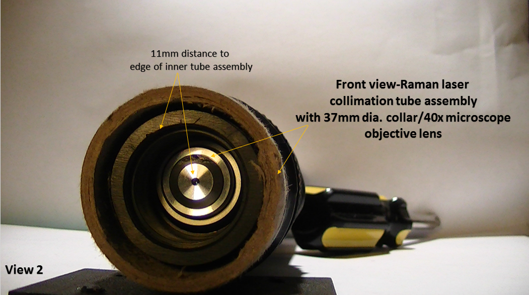
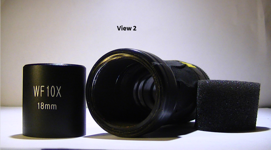
Raman Laser Collimation Tube Assembly/DAV5 V3 Spectrometer
12/28/2016 at 13:05 • 0 comments![]()
![]()
![]()
![]()
![]()
![]()
![]()
![]()
-
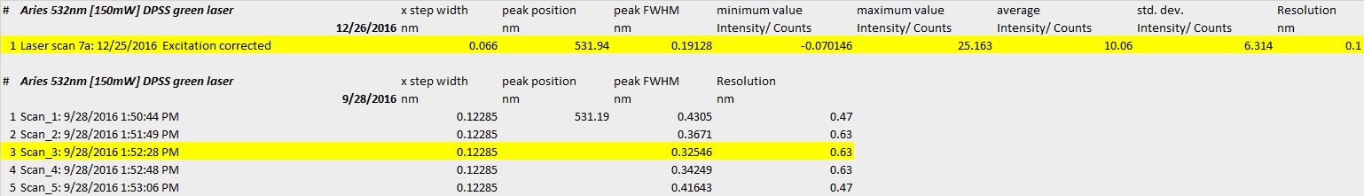
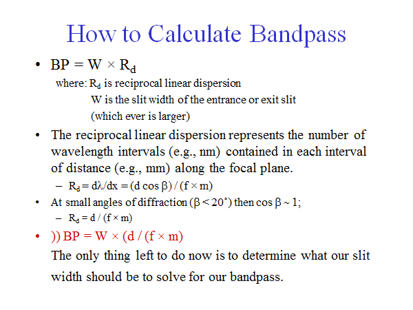
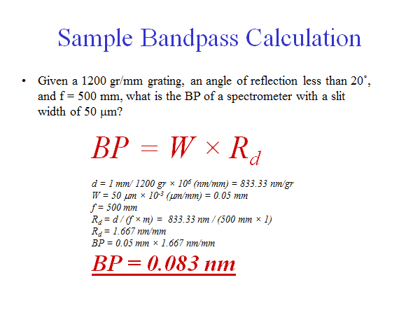
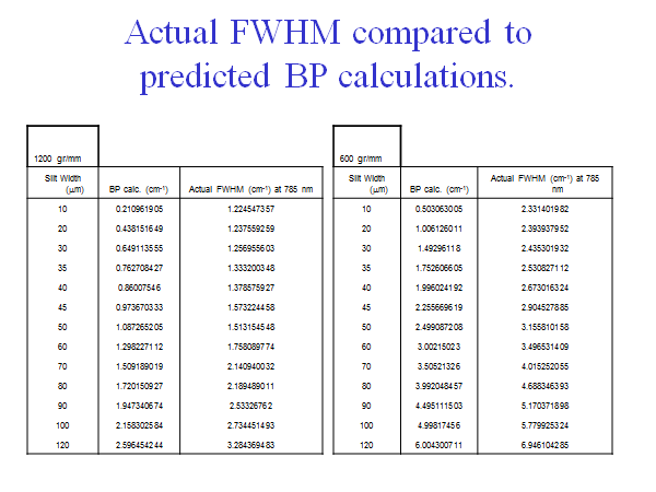
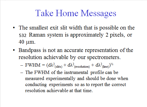
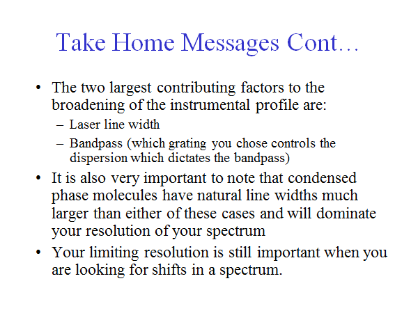
How Do You Calculate The FWHM of the Instrument Profile?
12/26/2016 at 22:55 • 0 comments![]()
![]()
Below is a comparison between a laser scan today (12/26/2016,) and one done on 9/28/2016
![]()
![]()
![]()
![]()
![]()
![]()
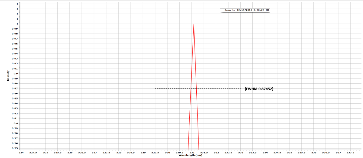
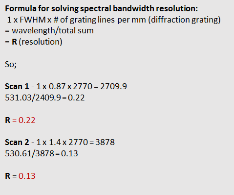
DAV5 V3 spectrometer; DVD diffraction grating-2770 gr/mm { 1 x 0.19 x 2770 = 5263}{531.94/5263 =0.10}
Resolution profile (present) = 0.10nm With a slit width of; 120um
![]()
![]()
What laser wavelengths are used for Raman spectroscopy?
Laser wavelengths ranging from ultra-violet through visible to near infra-red can be used for Raman spectroscopy. Typical examples include (but are not limited to):
- Ultra-violet: 244 nm, 257 nm, 325 nm, 364 nm
- Visible: 457 nm, 473 nm, 488 nm, 514 nm, 532 nm, 633 nm, 660 nm
- Near infra-red: 785 nm, 830 nm, 980 nm, 1064 nm
The choice of laser wavelength has an important impact on experimental capabilities:
- Sensitivity. Raman scattering intensity is proportional to λ-4 where λ is the laser wavelength. Thus an infra-red laser results in a decrease in scattering intensity by a factor of 15 or more, when compared with blue/green visible lasers.
- Spatial resolution. The diffraction limited laser spot diameter can be calculated according to the equation, diameter = 1.22 λ / NA (where λ is the wavelength of the laser, and NA is the numerical aperture of the microscope objective being used). For example, with a 532 nm laser, and a 0.90/100x objective, the theoretical spot diameter will be 0.72 µm – with the same objective, a 785 nm laser would yield a theoretical spot diameter of 1.1 µm. Thus, achievable spatial resolution is partially dependent on choice of laser.
- Optimisation of resulting based on sample behaviour. For example:
- Blue or green lasers can be good for inorganic materials and resonance Raman experiments (e.g., for carbon nanotubes and other carbon materials) and surface enhanced Raman scattering (SERS).
- Red or near infra-red (660-830 nm) are good for fluorescence suppression.
- Ultra-violet lasers for resonance Raman on bio-molecules (such as proteins, DNA, and RNA), and fluorescence suppression.
-
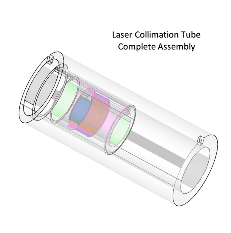
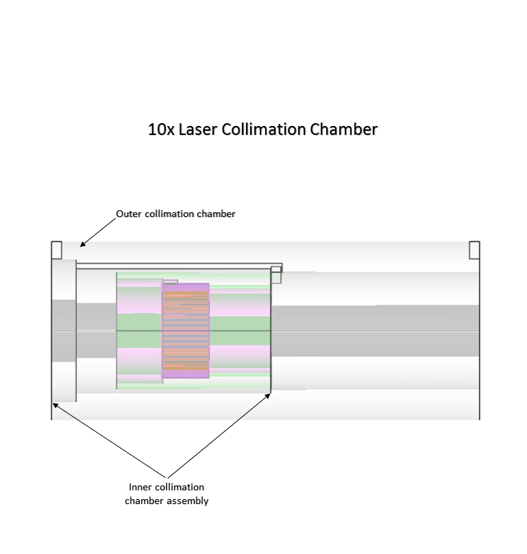
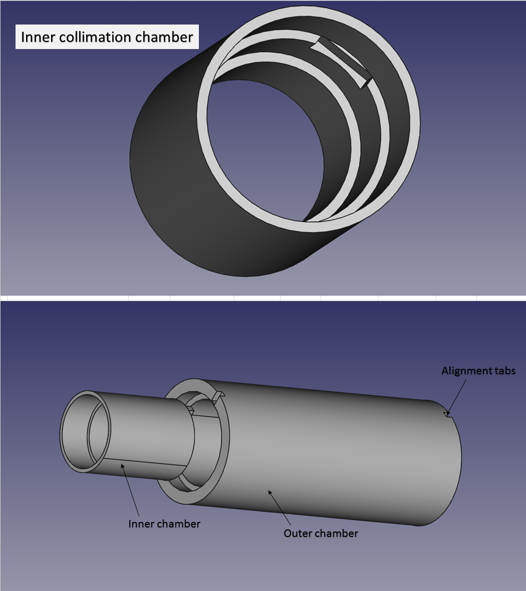
Section II Laser Collimation Tube/Prototype design for the DAV5 V3 spectrometer
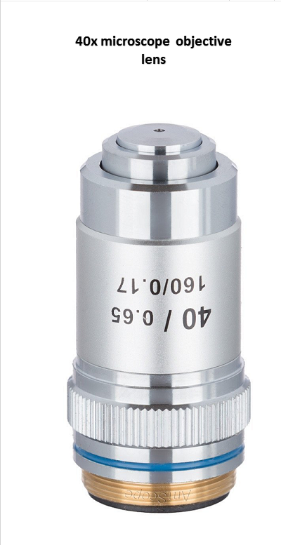
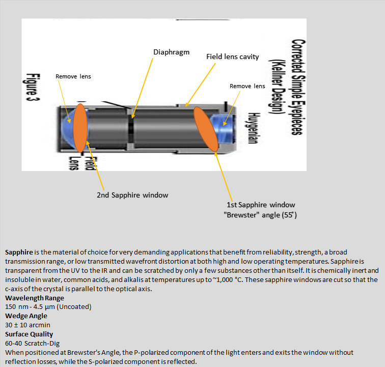
12/22/2016 at 22:04 • 0 commentsI used this illustration because this is the objective eye piece I used for the inner chamber of the collimation tube
![]()
![]()
![]()
![]()
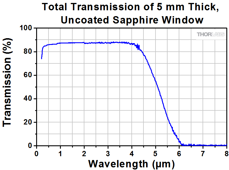
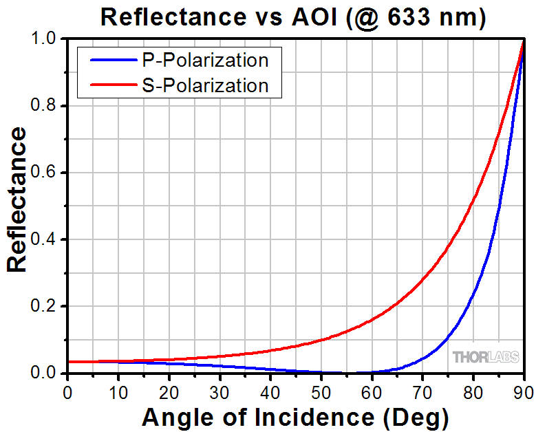
The graph below is the angle of incidence for the "brewster" angle for the 1st sapphire wndow:
![]()
![]()
![]()
![]()
![]()
![]()
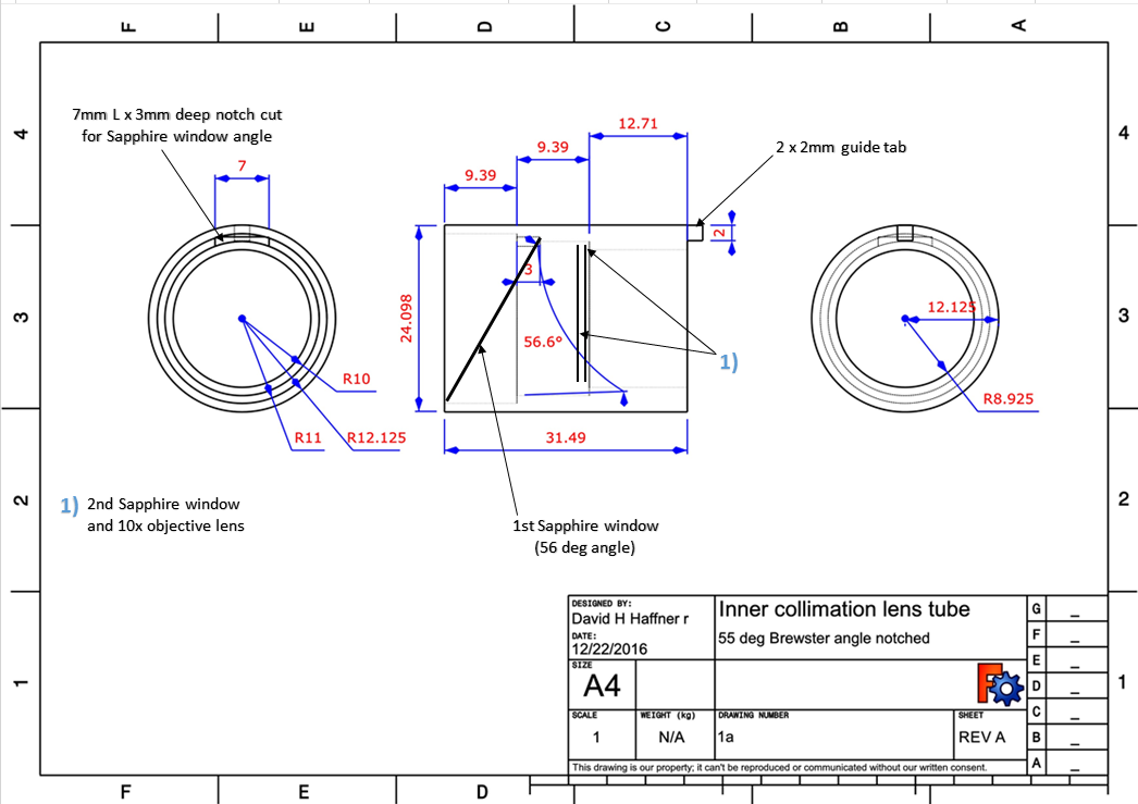
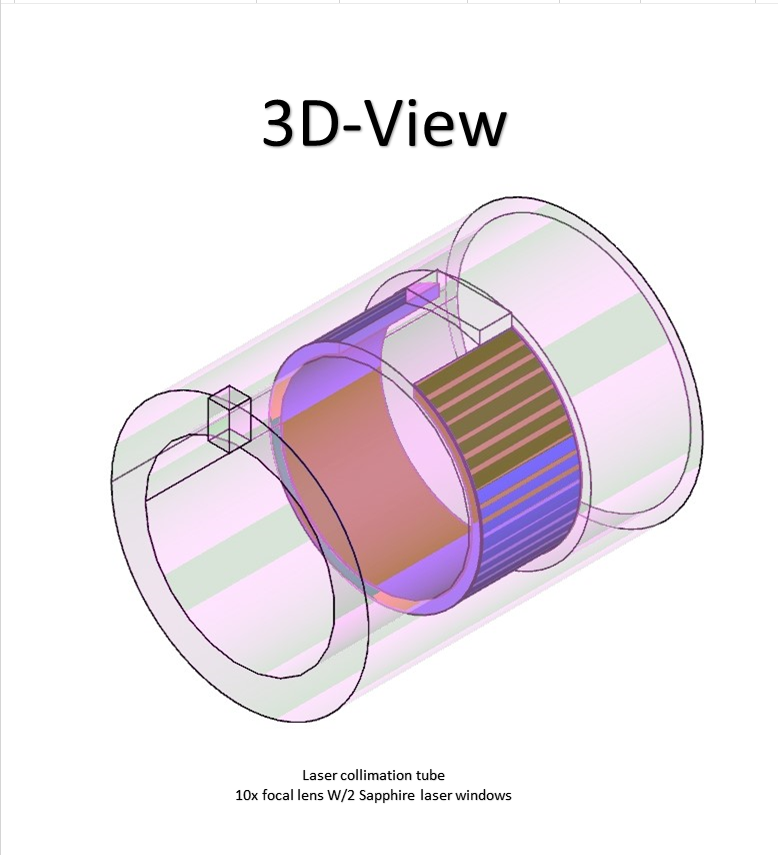
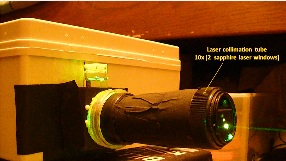
These parts have been uploaded as STL (mesh) files added to the components section, this is a critical piece of instrumentation, as the laser beam needs to be squeezed in very tightly and very precisely. The 2 sapphire windows, provide this accuracy and transmission, with no reflective aberrations occurring in the inner chamber.
-
Laser Collimation Tube/Prototype design for the DAV5 V3 spectrometer/Section I
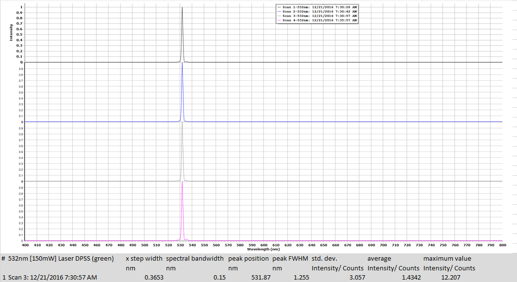
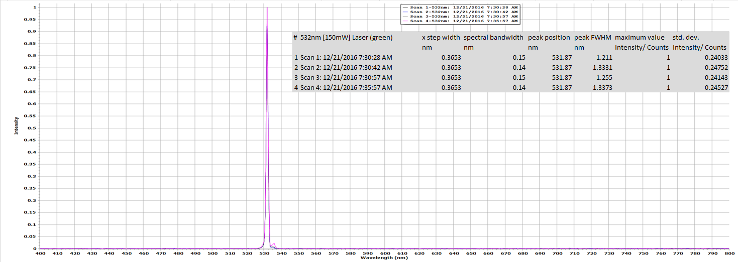
12/21/2016 at 22:26 • 0 commentsThis is my prototype laser Collimation tube for this spectrometer, this piece was very difficult to make with the precision that I had to make it in, but I did it. This is section (1) the initial data results using this design, very good results by the way! Section II will have the blueprints on how I made it, I just didn't want to take it back apart to take internal pics because it would have been too much of a problem to put it back together.
I will present those pictures and drawings in my next project log and how to assemble it.
![]()
![]()
![]()
![]()
![]()
![]()
-
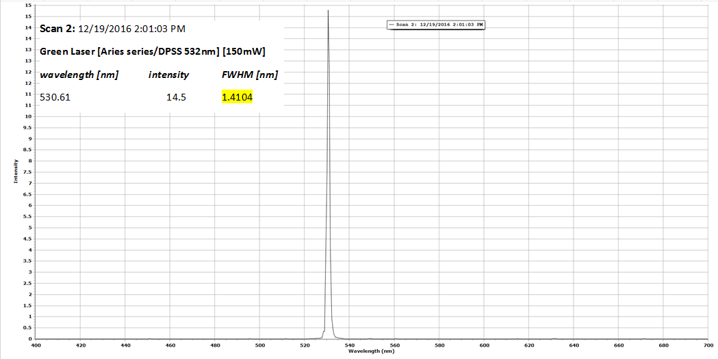
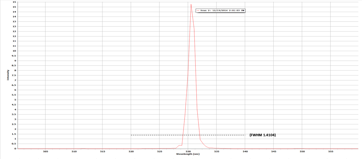
Laser Calibration of the DAV5 V3 Raman Spectrometer
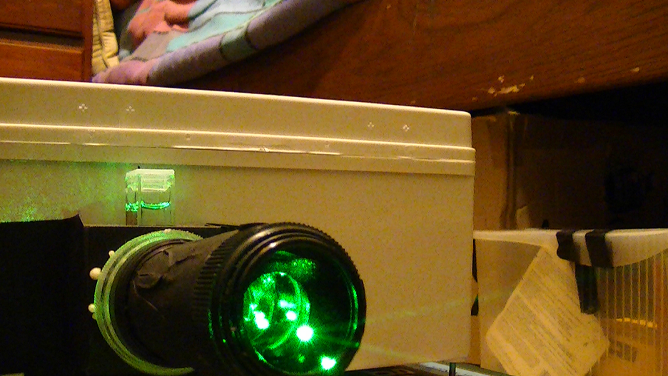
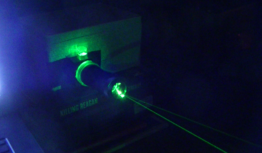
12/19/2016 at 21:18 • 0 comments12/19/2016; First serious calibration of the alignment and focus between the mirror, focal lens assembly and detector. I used a Quartz cuvette (clear on all 4 sides) this time, which has a wavelength range of 220nm - 2200nm and my Aries green 532nm DPSS high power laser (150mW,) I used a 99.99 % lab grade purity solution of Isopropyl alcohol in the quartz cuvette because its refractive index is 1.3772, so I would have only a crisp clear image of the laser line (spectra has been "normalized"):
![]()
![]()
![]()
![]()
![]()
References;
http://www.analiticaweb.com.br/newsletter/02/AN51721_UV.pdf
http://clinchem.aaccjnls.org/content/clinchem/21/11/1582.full.pdf
-
Fixed 32mm Focusing Lens Assembly for The DAV5 V3 Raman Spectrometer
12/19/2016 at 13:27 • 0 commentsI had to add a 5mm wide spacer behind the 32mm lens, I found through some experimentation that the increased 5mm distance sharpened the image significantly by a factor of 4, so the old focal distance was 65.2mm from lens apex to mirror's center, now it is 70.2mm from lens apex to mirror's center.
![]()
![]()
![]()
-
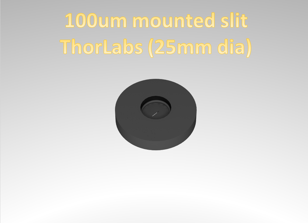
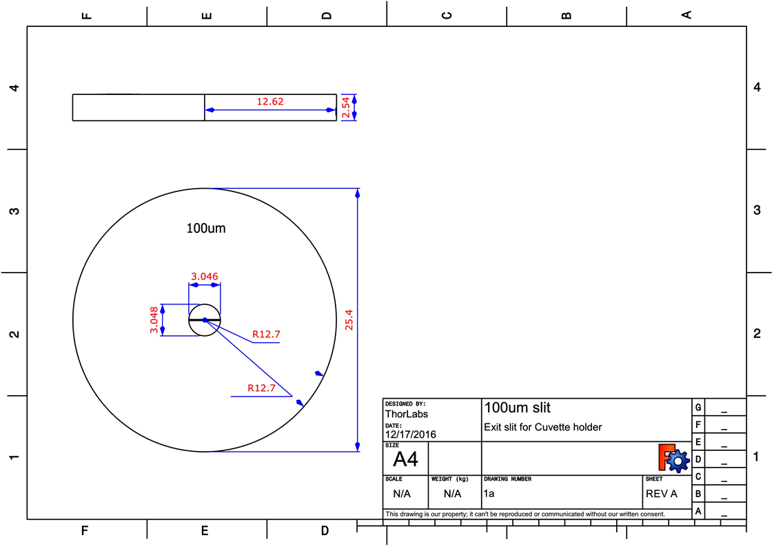
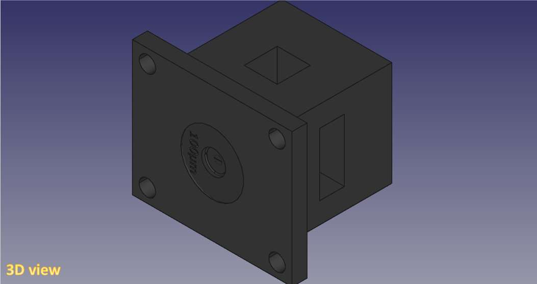
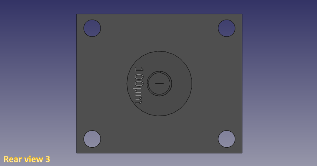
Re-designed Cuvette Holder W/100um exit slit mounted in rear plate
12/17/2016 at 14:43 • 0 commentsDAV5 V3 Raman spectrometer (prototype 1a)
![]()
![]()
![]()
![]()
![]()
DAV5 V3.01 Raman Spectrometer
The only thing worth doing, is the thing worth doing right!
 *Abstract, Before continuing any further in phase II testing, I wanted to see just where I am in both optical resolution and spatial resolving power. So I decided to take some spectral data from an EVOO sample I've had for awhile (preserved in a plastic UV cuvette W/an air tight top,) it's about 4 months old and some degradation does show up in the data but it was still an excellent sample to use.